Gruby (1843)
What is Microsporum?
Microsporum is a filamentous fungus belonging to the ecological group of pathogenic fungi called dermatophytes. Dermatophytes are keratinous fungi, which feed on protein keratin found in animal skin, nails, horns, and hair. They sometimes cause skin infections in humans and animals known as “ringworm”.

Like other dermatophytes, Microsporum belongs to the phylum Ascomycota, the family Arthrodermataceae. Along with the genera Trichophyton, Epidermophyton, and Chrysosporium, the genus Microsporum comprises of asexual stages. The sexual stages belong to the genus Arthroderma [1].
Like other dermatophytes, Microsporum species can live saprophytically in soil, where they derive nutrients from substrates such as hair, feathers, and horns present in the environment. People and animals can become infected with fungi through contact with contaminated soil. Certain Microsporum species have specific animal hosts, while others can infect humans too [1].
How many species of Microsporum are there?
The genus Microsporum consists of eighteen species, with clinically important being M. canis, M. gypseum, M. gallinae, M. nanum, and M. audouinii [1]. Some Microsporum species are ubiquitous, and some are endemic, colonizing only specific geographical regions.
- M. canis is the major infectious agent of tinea diseases in Europe, the eastern Mediterranean, South America, Asia, and Africa. It is considered the predominant species that are causing skin diseases in cats and dogs. In cattle, it can cause bovine ringworm [1]. In humans, it causes capitis (fungal infection of the scalp) and tinea corporis (fungal infection of mostly arms and legs).
- M. audouinii causes tinea capitis in children in North America, Asia, and Africa.
- M. gypseum can survive in the environment absent of keratinous substrates by colonizing substrates such as sewage sludge and cave soils. It was isolated from human, feline, and canine dermatophytic lesions in Asia and Africa. M. gypseum is also reported as an infectious agent of ringworm in cattle and horses [1].
- M. nanum, along with M. canis and M. gypseum, is the causative agent of dermatitis in pigs, while M. gallinae causes favus skin disease (white comb) in birds [1].
What does Microsporum spp. look like?
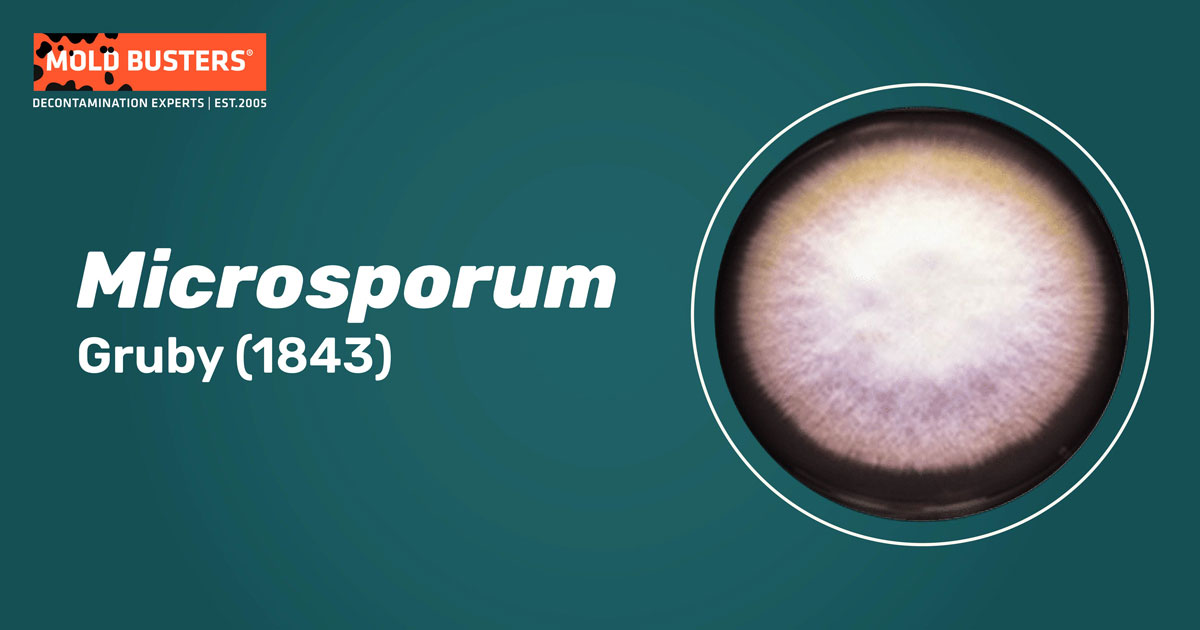
Microsporum spp. form large velvety or powdery colonies with variable pigmentation. While M. canis produces lemon yellow colonies, colonies of M. nanum are rust-colored [1]. In the environment and laboratory cultures, Microsporum species produce septated branching hyphae. As an asexual fungal stage, Microsporum species reproduce by asexual spores – conidia.
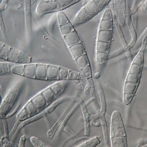
There are two types of conidia: macroconidia and microconidia. Macroconidia are arranged along the hyphae and can usually be very large (40–150 μm in length and 8–15μm wide). They are spindle-shaped to ovoid, rough-walled, and divided by septa (Fig. 1). The powdery surface that mature colonies display is due to the abundant production of macroconidia. Microconidia are stalked or sessile, club-shaped, usually 1.7–3.5μm wide x 3.3–8.3μm long. They are formed singly along the hyphae [1, 4].

What is Microsporum infection?
The infectious agent of these species is chlamydospores or arthrospores. These thick-walled spores are produced vegetatively from mycelial cells and are resistant to unfavorable conditions. They can survive in the environment for several years and germinate when appropriate conditions arise [1, 2].
Since spores cannot invade the healthy skin of people or animals, they have to be introduced into host tissue by injury (wound pathogens) or through vector organisms such as cats or dogs. After penetration through injured skin, spores of Microsporum spp. germinate into the epidermis, and the hyphae grow along with the hair [1].
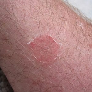
Around hair follicles, hyphae produce spores that remain attached to the hair shaft by forming a thick coating. Microsporum cannot colonize deeper layers of the skin, so hair grows normally. However, some hair breaks near the skin surface and causes alopecia. Fungal metabolites can cause inflammatory reactions at the site of infection, and this enables the pathogen to move away from the site of infection, forming the circle. The typical ringed lesion (Fig. 2) caused by Microsporum sp. is characterized by healing in the center and papules on the periphery [1].

Feline ringworm caused by M. canis is characterized by regular, circular alopecia, 3-6 cm (1.18-2.36 inches) in diameter. Lesions are single or multiple, with central healing and inflamed, scaly skin at the margin of circles. Lesions are typically present on the head but can also be found on the legs and tail [1]. Immunosuppressed cats can also develop ear infections characterized by a waxy discharge from the ear canal. Dog ringworm lesions are commonly seen on the faces, paws, and elbows of infected animals. Cats and dogs are considered reservoirs of M. canis [2].
Like in animals, M. canis infection in humans has been associated with alopecia, scaling, and circular lesions in localized forms, such as tinea capitis (scalp infection), tinea corporis (body infection), tinea pedis (foot infection), and onychomycosis (nail infection) [2].
Tinea capitis is the most frequent human infection caused by M. canis and M. audouinii. It commonly occurs in children that have been in close contact with infected animals. Symptoms are mild inflammation and patchy areas of scaling. In immunocompromised children, inflammation can become more serious, followed by alopecia. M. canis and M. gypseum can cause progressive mycosis of deeper layers of the skin scalp, causing the formation of tumorous masses with crusts [1]. Human onychomycosis is a nail infection caused by M. canis, and it is very rare since Microsporum generally does not infect the nails [1].
Tinea incognito is a condition in humans caused by M. canis, resulting from poor diagnosis and prolonged corticosteroid application [1, 3, 5]. Symptoms include plaques with inflammation and papules on the neck and face, followed by hair loss.
How to treat M. canis?
In animals, weak to moderate infections can often be resolved without medical intervention. However, treatment is recommended to prevent transmission to humans [1]. Due to poor penetration of antifungals through the hair coat, topical application is not effective. Systemic treatment of infected animals includes itraconazole administered by pulse therapy. Other antimycotics such as terbinafine, ketoconazole, and griseofulvin are effective but not recommended due to their adverse effects on animal health. Vaccines based on M. canis are ineffective since they produce poor immunity against this pathogenic fungus [1]. Weekly applications of lime sulfur, enilconazole, or a miconazole/chlorhexidine shampoo are currently recommended for decontamination of animal fur [2].
In humans, treatment of tinea capitis due to M. canis infection commonly includes oral application of antifungals griseofulvin, terbinafine, and itraconazole [1]. In extensive infections, systemic treatment should be supported by using topical antifungal and antibacterial therapy [1, 2].
Since M. canis infections can be very transmissible, treatment methods are required to avoid potential transfer to additional susceptible hosts. Treatment failure has been reported in 25–40% of treated patients due to a lack of patient compliance, drug penetration into tissue, medication bioavailability, and resistance phenomena [2]. When it comes to dermatophytosis, it is important to precisely identify the causal agent and estimate appropriate treatment, avoiding the development of tinea incognito and further complication [3].

Did you know?
The #1 toxic mold type found in homes is the Penicillium/Aspergillus mold group?! Find out more exciting mold stats and facts inside our mold statistics page.
References
- Samanta, I. (2015). Veterinary Mycology. Springer, New Delhi, India. pp 21-29.
- Aneke, C. I. et al. (2018). Therapy and Antifungal Susceptibility Profile of Microsporum canis. Journal of Fungi. 4 (3): 107
- Rodríguez, P. A. et al. (2018). Human dermatophytosis acquired from pets: report of three cases. Journal of Dermatology and Cosmetolo 2(4):38-41.
- Howard, D. H. (Ed.). (2002). Pathogenic fungi in humans and animals. CRC Press.
- Romano, C., Asta, F., & Massai, L. (2000). Tinea incognito due to Microsporum gypseum in three children. Pediatric dermatology, 17(1), 41-44.

Get Special Gift: Industry-Standard Mold Removal Guidelines
Download the industry-standard guidelines that Mold Busters use in their own mold removal services, including news, tips and special offers:

Written by:
Jelena Somborski
Mycologist
Mold Busters
Edited by:
Dusan Sadikovic
Mycologist – MSc, PhD
Mold Busters
