Myxomycetes, or slime molds, are a group of free-living amoeboid and sessile primitive organisms with complicated life cycles.
Despite not belonging to the kingdom of fungi, they were historically regarded as molds, due to the similarities in appearance and lifestyle.
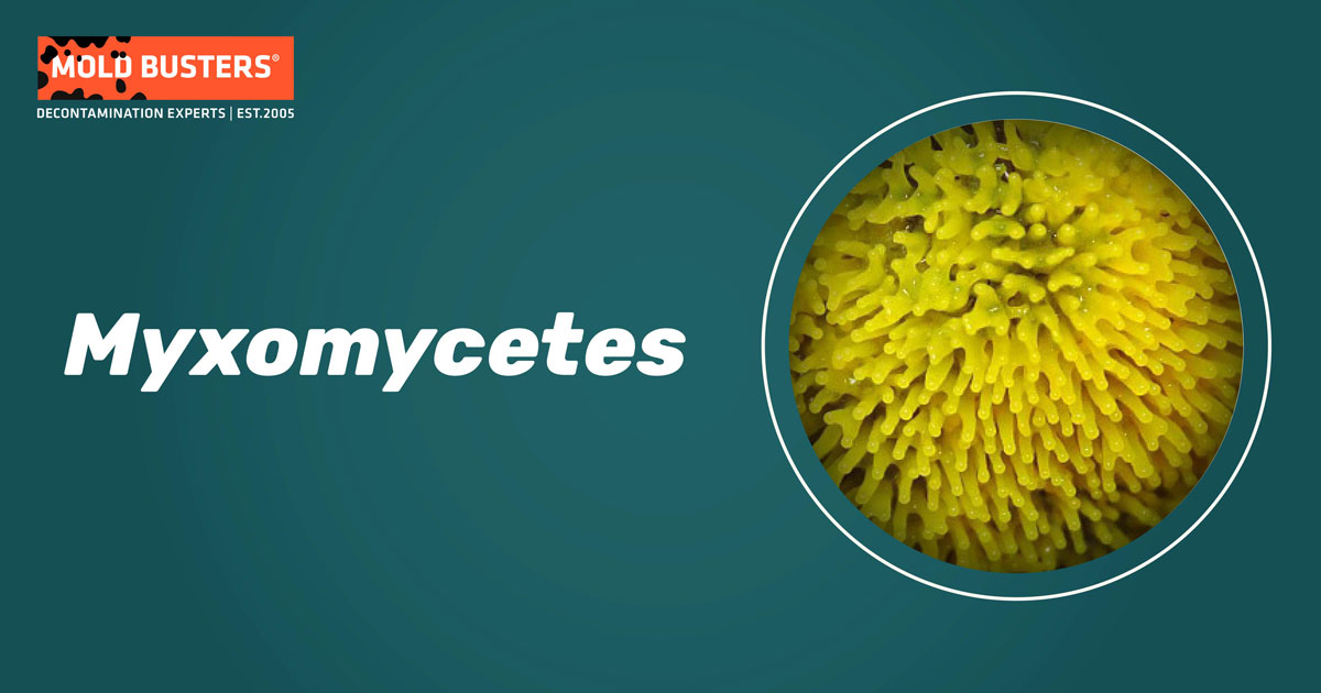
Unlike mostly saprotrophic fungi, the Myxomycetes are free-living predators of bacteria and eukaryotic protists.
Why are Myxomycetes called slime molds?
Throughout history, the Myxomycetes have been variously classified as plants, animals, or fungi. Due to similarities to true molds (shared habitats and reproduction via aerially dispersed spores), Myxomycetes have traditionally been studied by mycology.
The name most closely associated with the group, originally used by the German naturalist Link nearly 200 years ago, is derived from the Greek words “myxa“ (mucus or slime) and “mycetes” (referring to fungi) [1]. However, abundant molecular evidence now confirms that they are amoebozoans and not fungi [2].
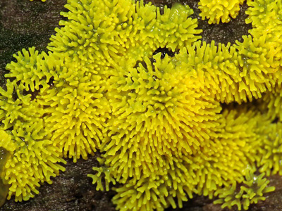
Furthermore, even though the idea of slime molds being a different group of organisms was pointed out during the 1800s, Myxomycetes continued to be considered as fungi by most mycologists until the second half of the twentieth century.

What are slime molds?
Although they have the word ‘mold’ in their common name, Myxomycetes aren’t fungi. Unlike true fungi, they live a predatory lifestyle, do not form mycorrhizae, and have no role in the decomposition of lignocellulosic matter. There are approximately 1000 species of slime mold [1].
The life cycle of slime molds consists of two distinct stages. During the amoeba-flagellate stage, slime molds exist as typical single-celled organisms. These amoeboid cells feed on bacteria, grow and multiply by binary fission. This stage eventually progresses to the plasmodium stage, usually via the sexual fusion of two compatible amoeboflagellates.
Morphologically the plasmodium is a veiny network with a viscous, slimy consistency. Most of the life cycle is spent in this stage. Plasmodia may or may not be visible to the naked eye and can be transparent, white, or brightly colored (which is more often the case). The plasmodium is multinucleate and can occupy an area larger than one square meter. However, it is technically still one cell, as it undergoes many nuclear divisions but no true cellular divisions. A specimen of Brefeldia maxima found in Wales weighed around 20 kg – one of the largest single-cell organisms ever recorded [3].
Unlike true fungi, the plasmodium feeds on bacteria, fungal hyphae, and other microorganisms, ingesting them through phagocytosis. Also, in contrast to fungi, the plasmodium has chemotactic and phototactic abilities, meaning it can move towards or away from chemical stimulants or light.
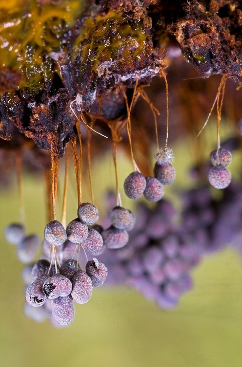
If the conditions are right, the plasmodium will begin to produce fruiting bodies. The plasmodia dry out and harden overwinter until the spring snow thawing, winds and participation release the spores and the entire cycle will start again. Slime molds fruiting bodies exhibit a wide variety of colors, shapes, and sizes. Considering the similarities between a plasmodium with fruiting bodies and a sporulating fungal mycelium, it is no wonder that the Myxomycetes were mistaken for fungi at first.
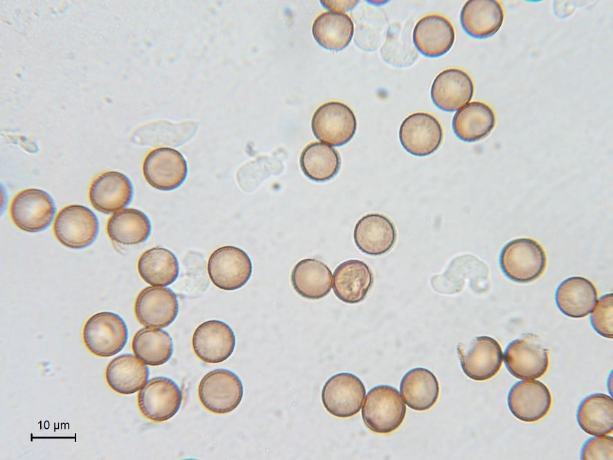
The spores of Myxomycetes are microscopic and light and can be carried by the wind for considerable distances. Their walls are generally patterned, containing reticular, warty, or spiky motifs, and are very rarely smooth (Fig. 3). Myxomycete spore size usually ranges between 5-20 µm large. However, these traits vary from species to species, and the color, shape, and spore diameter are important taxonomic characteristics. They can survive a wide range of adverse conditions, often remaining in a dormant state for prolonged periods. It is not uncommon for spores to germinate years or decades after they were first released. Their limiting conditions for germination are moisture and temperature.
Myxomycetes classification
Since their discovery, the classification of Myxomycetes has been revised many times. This is unsurprising considering the early confusion about their precise nature. In fact, 12 alternative classifications of Myxomycetes have been proposed since 1950 [6].
The recent phylogenetic analyses divide slime molds into two superclades depending on the appearance of their spores and further into several orders and families. However, researchers suggest that many classical taxa are paraphyletic (meaning that group members are from the same lineage but some descendants are missing) and that their classification needs major revision [7].
According to Stephenson and Schnittler [1] the classification of Myxomycetes can be summarized (to the level of order) as follows:
- Myxogastria
- Collumellidia
- Echinosteliales
- Physarales
- Stemonitales
- Lucisporidia
- Liceales
- Trichiales
- Collumellidia
Where can slime molds be found?
Myxomycetes have been observed in most terrestrial habitats [1]. They are common in forests, often developing on tree bark, decaying logs, stumps, dead leaves, and other organic litter. Similar to true molds, they are mostly associated with cool and moist habitats. Although most slime molds are terrestrial, several species have been reported from aquatic habitats. The plasmodia of these species live completely underwater and only emerge to develop fruiting bodies [4].
Most species have a cosmopolitan distribution. However, some species can only be found in the tropics or only in temperate regions. Interestingly, the diversity of slime molds seems to be greater in temperate regions than in the tropics [4]. Experts believe that this is due to several reasons, notably high amounts of rainfall that wash away different stages of slime mold development, a lack of air movement that is detrimental to spore dispersal. Furthermore, high humidity promotes fungal invasion. However, it has also been suggested that the Myxomycetes that occur in tropical forests only occasionally form fruiting bodies, thus evading detection in these already poorly investigated habitats.
Some species of slime molds (referred to as strictly nivicolous species) that live at the high altitudes of the Rocky Mountains and the Alps are often found fruiting on melting snowbanks in late spring and early summer. Also, some species occur in Arctic and Antarctic environments and have been collected from places such as Alaska, Iceland, Greenland, and the Antarctic Peninsula [4]. Additionally, several species have been reported in extremely dry places such as the Atacama Desert, parts of the Arabian Peninsula, and in western Mongolia [1].
Although slime molds are not generally associated with indoor environments, they likely exist in our houses in one shape or another. One study sampled leaves and stems of common house plants revealed that 70% of the cultures showed the development of slime molds, mostly belonging to the Physarales order [5]. Therefore, it is reasonable to assume that slime molds are ubiquitous, and possibly in our homes, although remaining in dormant states for the most part.
Myxomycetes mold statistics
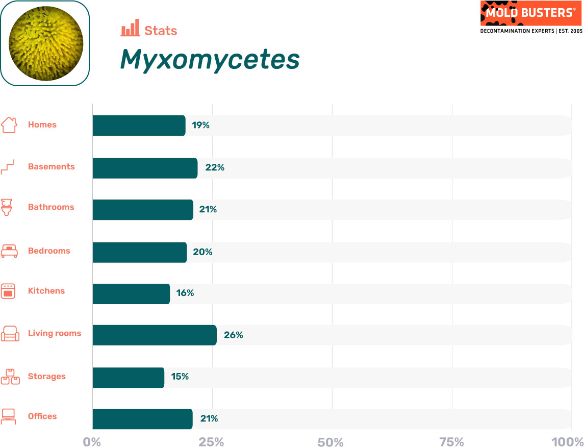
As part of the data analysis presented inside our mold statistics resource page, we have calculated how often mold spore types appear in different parts of the indoor environment when mold levels are elevated. Below are the stats for Myxomycetes:

Are Myxomycetes dangerous?
Myxomycetes are not known to be pathogenic or of economic importance. Due to these facts, they were poorly studied. A recent study showed that 42% of subjects who suffer from seasonal allergic rhinitis had positive skin test reactions to myxomycete spores. Interestingly, several subjects who had negative allergy prick tests to standard aeroallergens showed sensitization to myxomycete spores, suggesting that Myxomycetes could account for some of the “unknown seasonal allergens” that are also thought to cause seasonal allergic rhinitis [8].
However, associations between Myxomycetes and humans are rare. Although several species are known to form fruiting bodies on living plants, including ornamentals, grasses, and crops, the damage to the plants is practically non-existent and the slime molds are washed off with the first rain.
Interactions between Myxomycetes and humans are so rare that most people don’t even know what they look like. A particularly large growth of Fuligo septica in a neighborhood of Dallas in 1973 caused quite an alarm and even made the nationwide news. The mass broke apart when washed away with water, but the blobs continued to crawl about and increase in size, causing panic amongst citizens. Eventually, the blobs were identified as nothing more than a slime mold and the excitement waned [4].
In any case, slime molds are usually nothing to worry about. However, if you have any questions about slime molds or any true fungal species, feel free to call Mold Busters today!
References
- Stephenson SL, Schnittler M (2017). Myxomycetes. In: Handbook of the Protists (Second Edition). Springer International Publishing. pp 1404-24.
- Yoon HS, Grant J, Tekle YI, Wu M, Chaon BC, Cole JC, Logsdon JM Jr, Patterson DJ, Bhattacharya D, Katz LA (2008). Broadly sampled multigene trees of eukaryotes. BMC Evol Biol. 8:14.
- Ing B (1999). The Myxomycetes of Britain and Ireland : an identification handbook. Richmond Publishing Company. Slough, England. pp 4.
- Stephenson SL, Stempen H (2000). Myxomycetes: A Handbook of Slime Molds. Timber press, Inc. pp 49-58.
- Tucker DR, Coelho IL, Stephenson SL (2011). Household slime molds. Nova Hedwigia. 93(1-2):177-182.
- Wikispecies: Free Species Directroy (2019). Myxogastrea. Retrieved from wikimedia.org
- Adl SM, Simpson AG, Lane CE, Lukeš J, Bass D, Bowser SS, Brown MW, Burki F, Dunthorn M, Hampl V, Heiss A, Hoppenrath M, Lara E, Le Gall L, Lynn DH, McManus H, Mitchell EA, Mozley-Stanridge SE, Parfrey LW, Pawlowski J, Rueckert S, Shadwick L, Schoch CL, Smirnov A, Spiegel FW (2012). The revised classification of eukaryotes. J Eukaryot Microbiol. 59(5):429-93.
- Lierl MB (2013). Myxomycete (slime mold) spores: unrecognized aeroallergens? Ann Allergy Asthma Immunol. 111(6):537-41.e2.